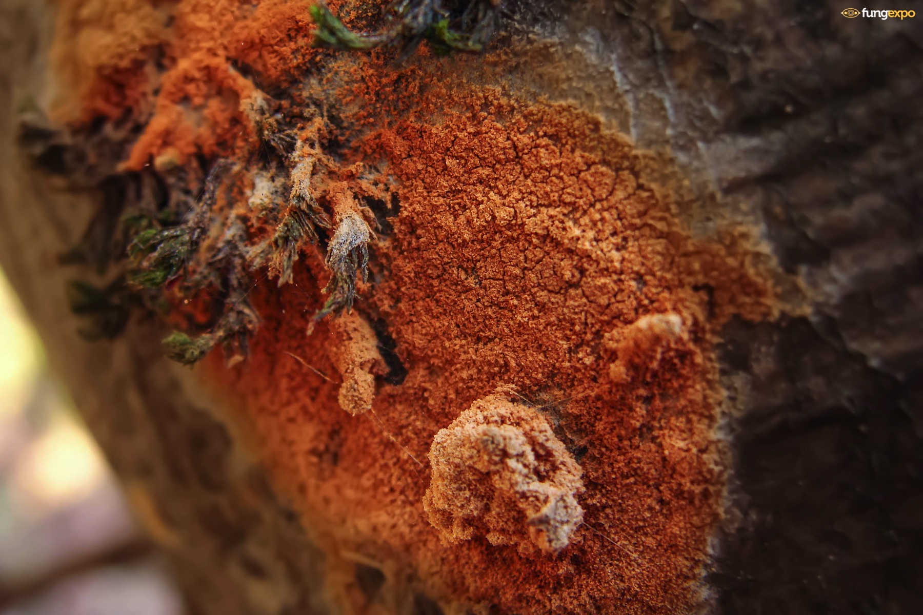
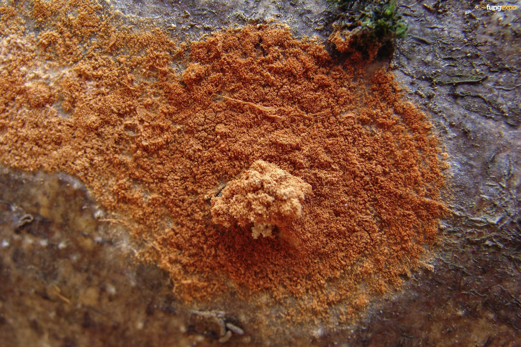
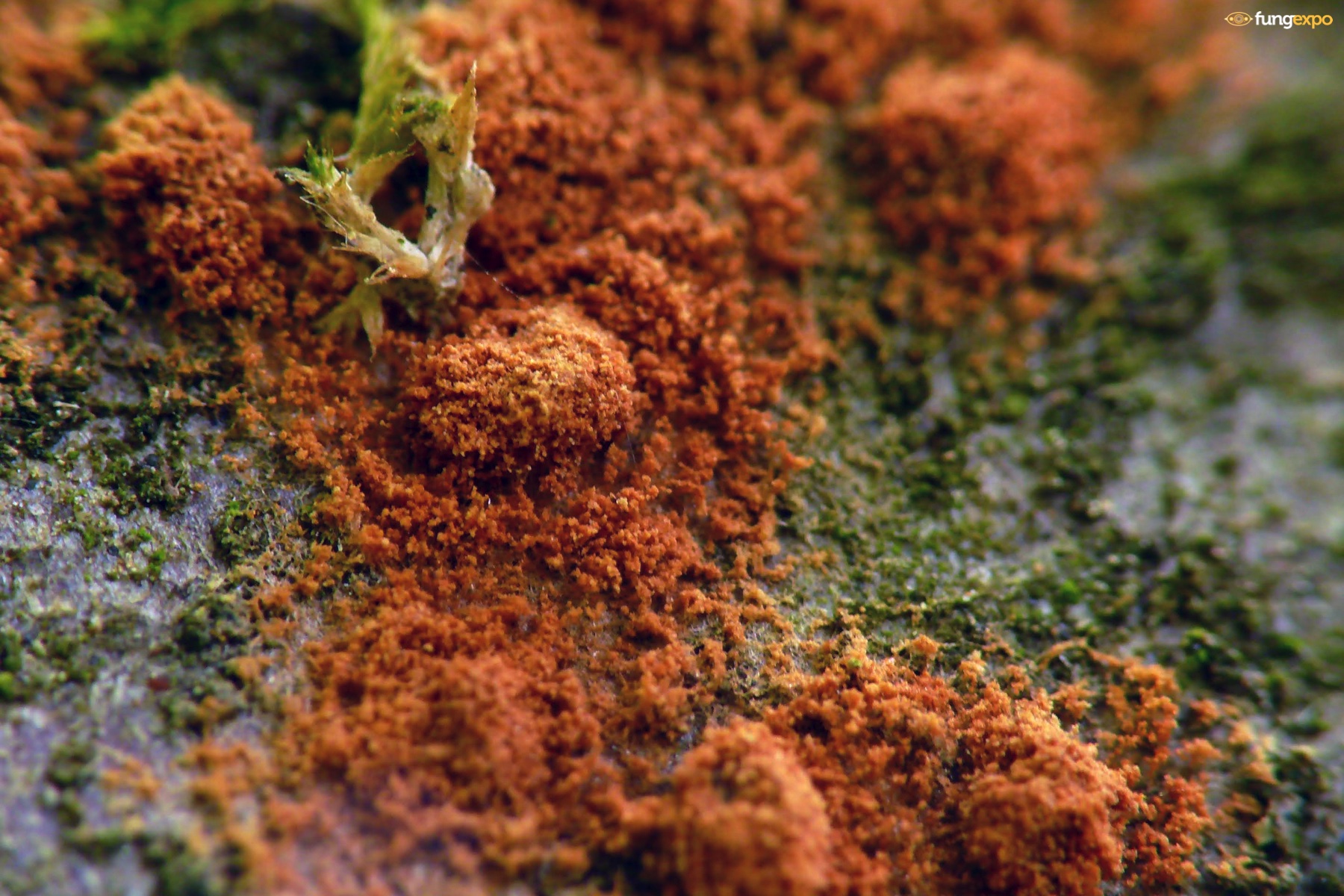
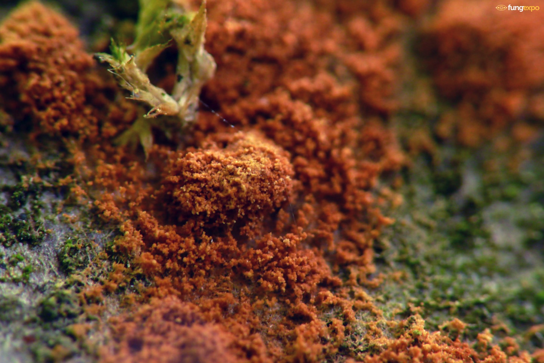
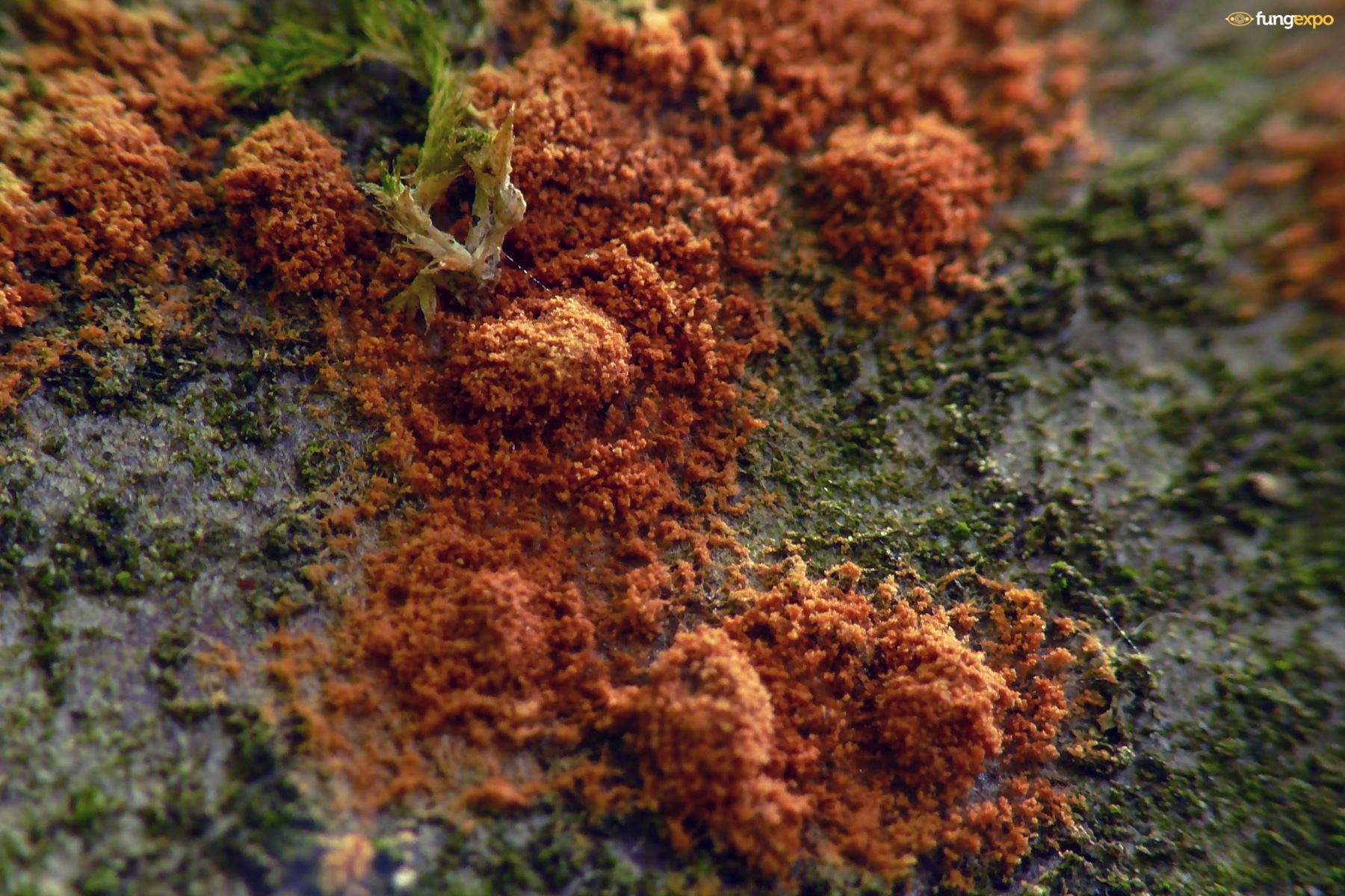
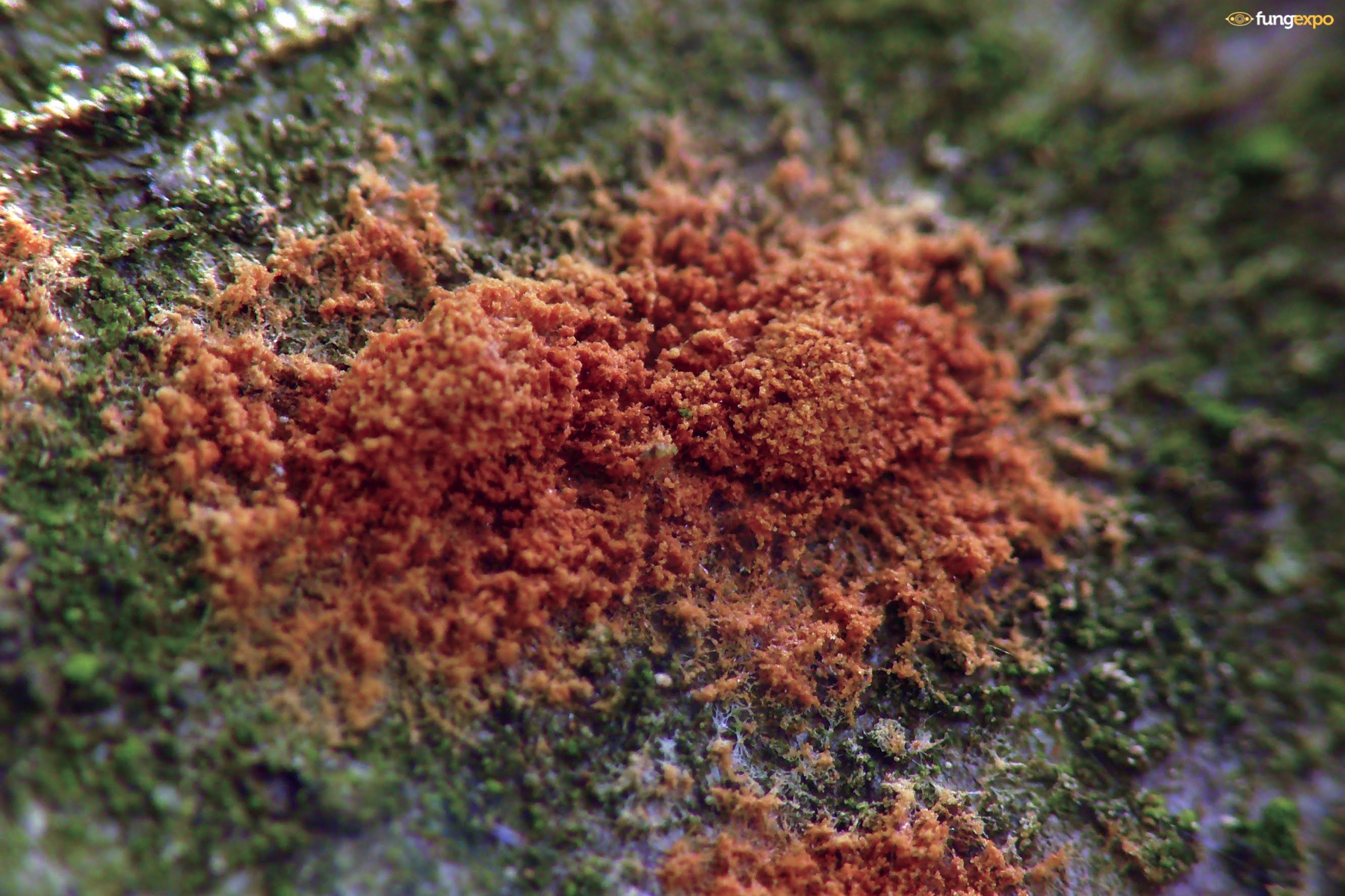
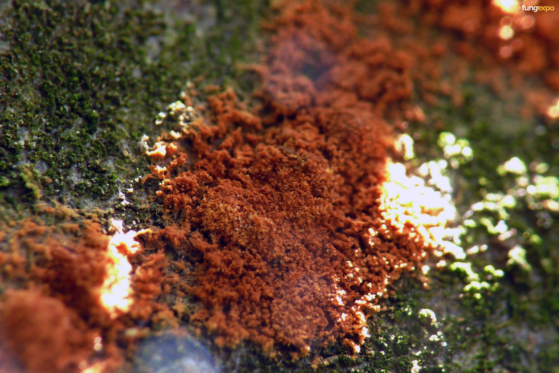
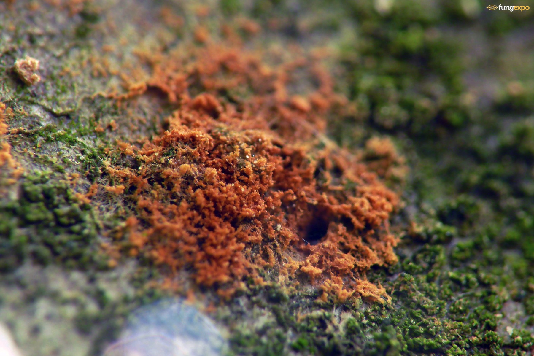
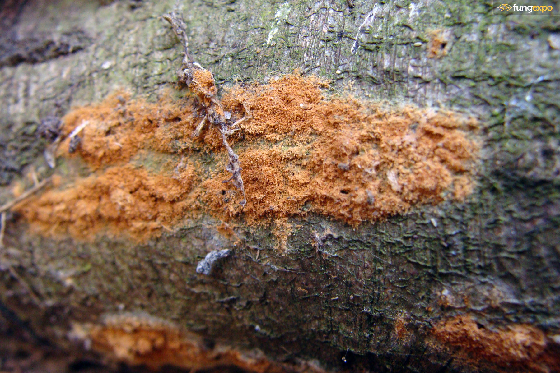
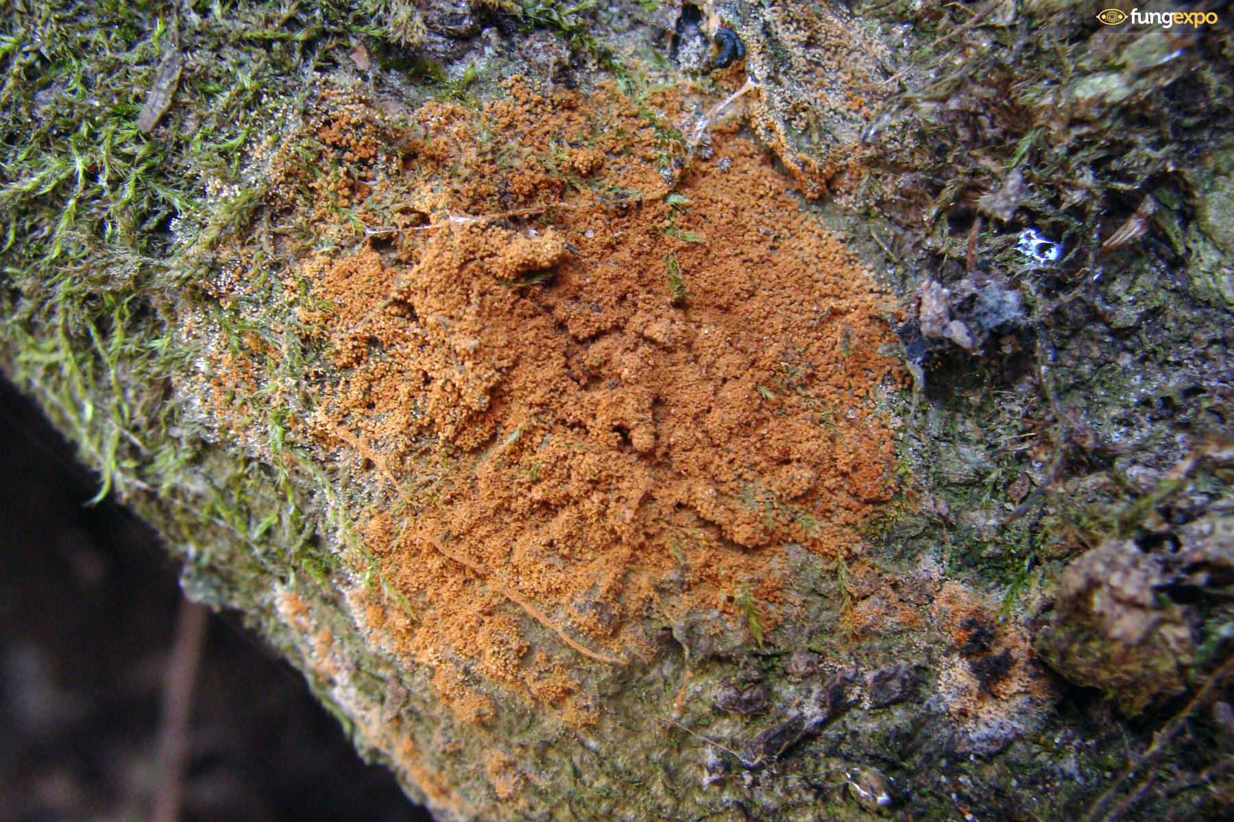
In situ photos:
Microscope Photos:
A 2017.06.03. Ω
In situ photos:
Microscope Photos:
 Notes:
2024. January 10., BG
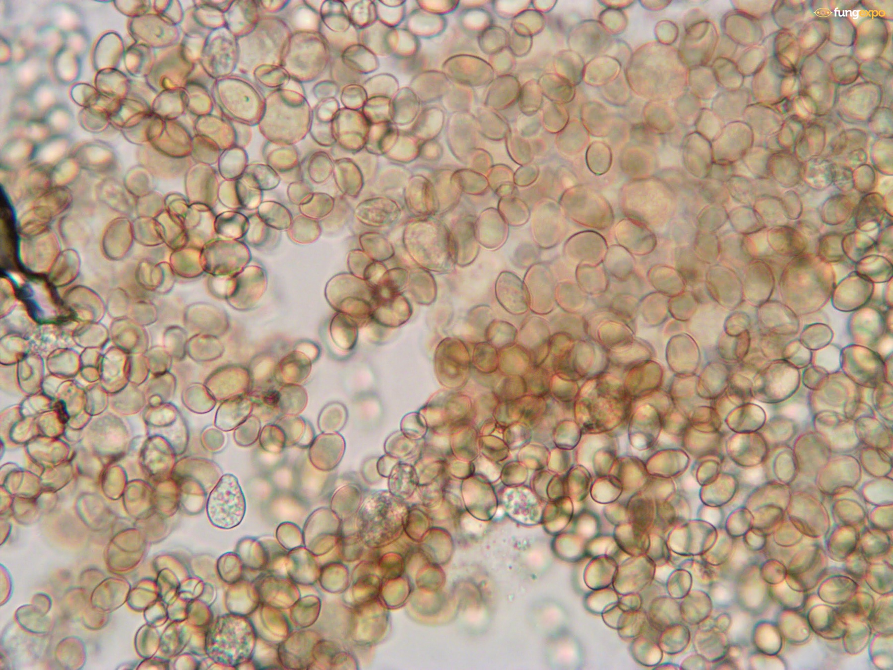
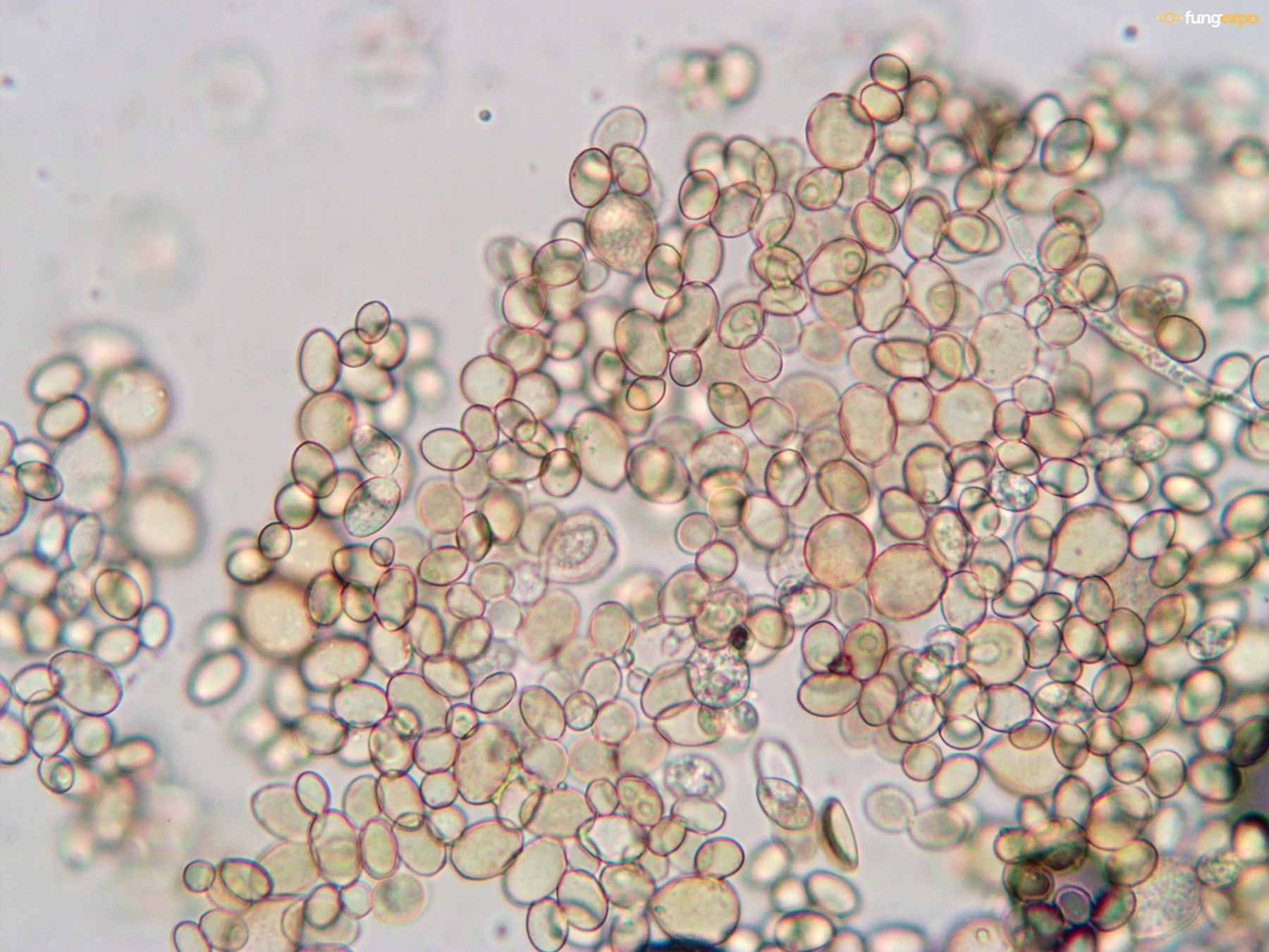
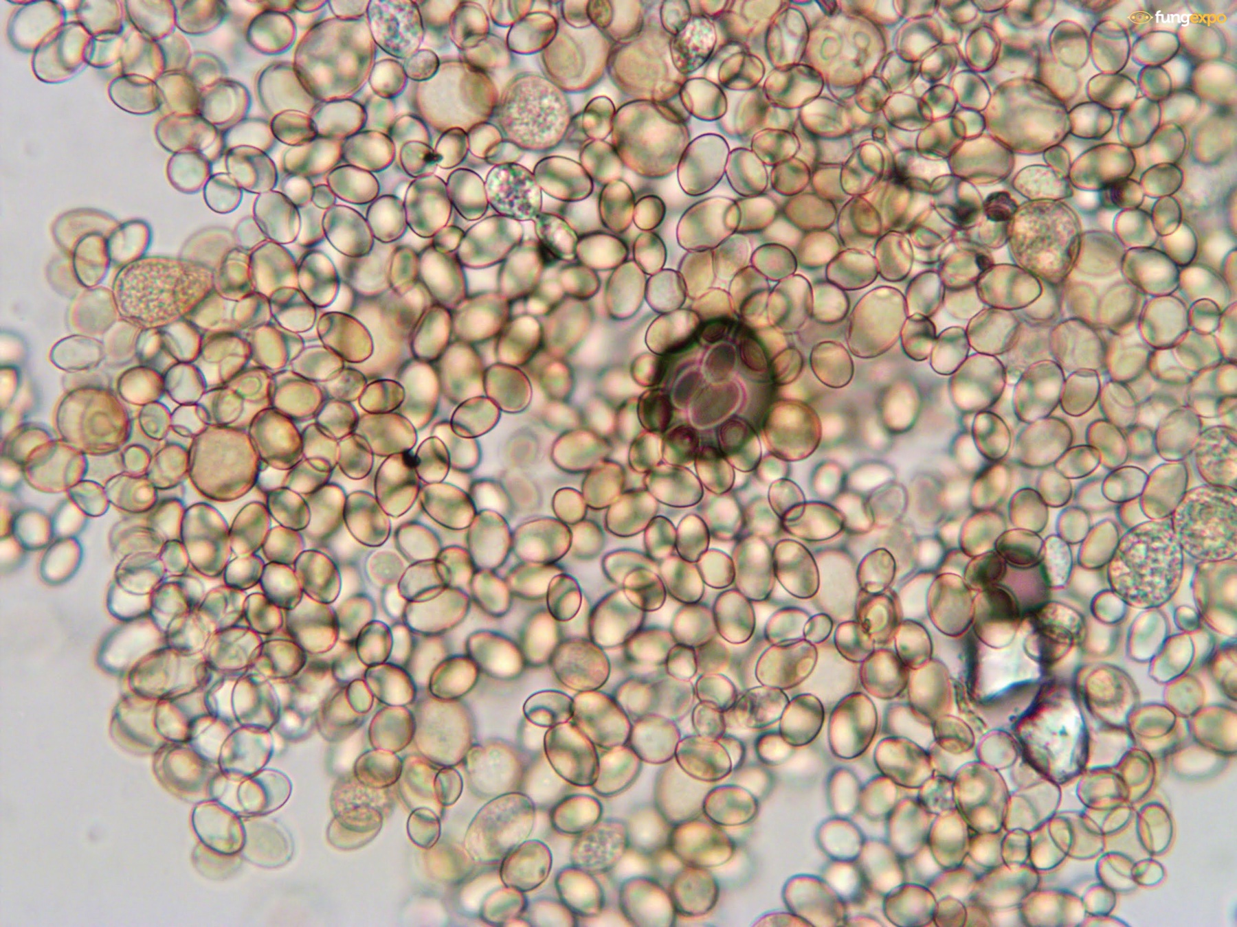
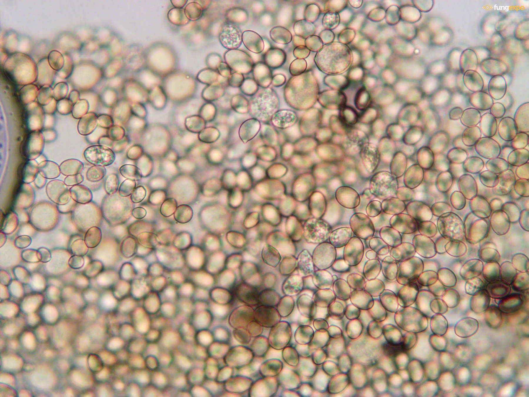
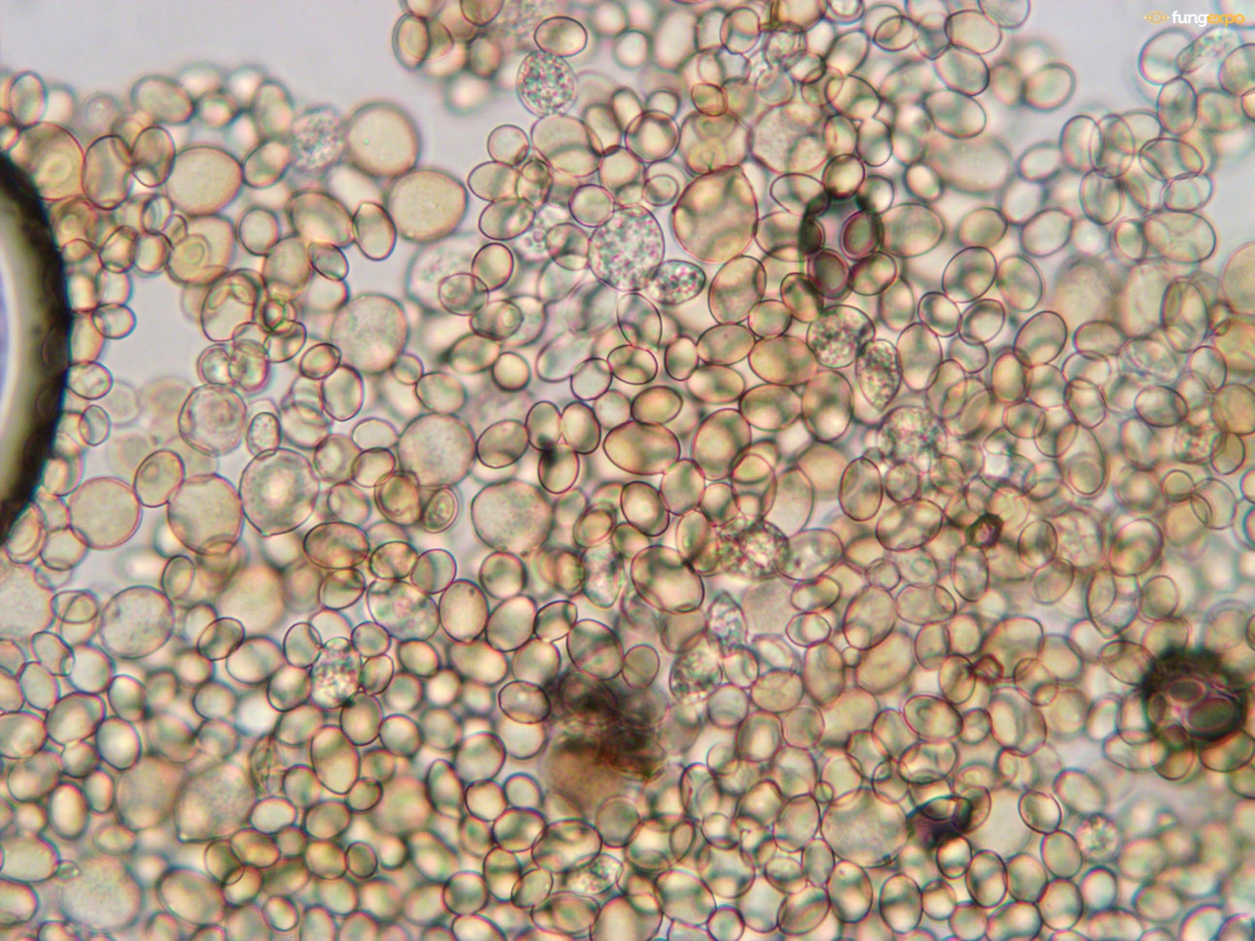
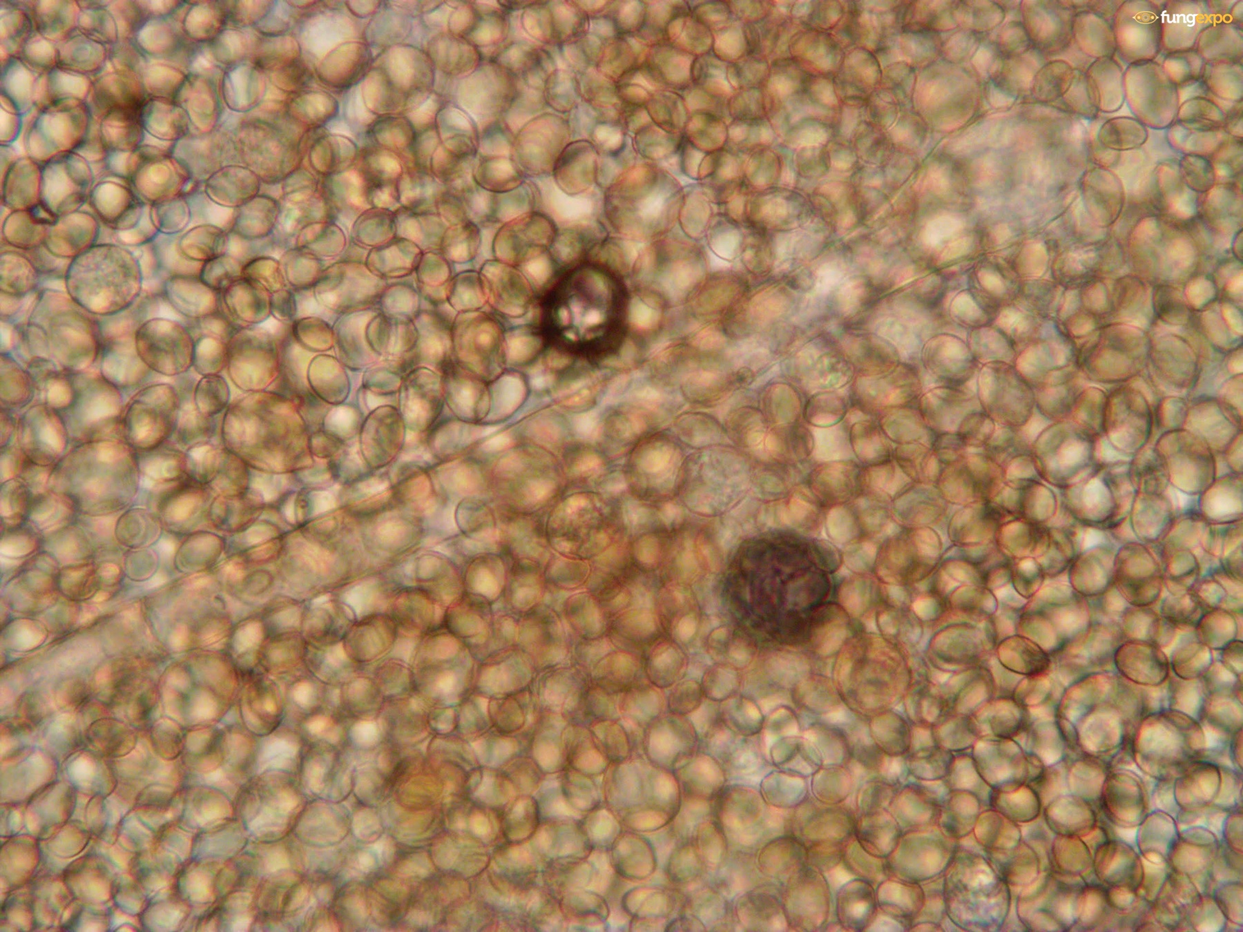
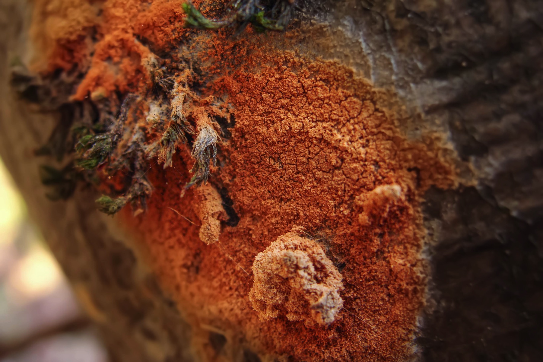
Mikromorfohorsegiailag elég komplex felépítményű ez a mikoparasite gomba, a fungexpo gyorsleírásai ehhez nem tudnak kielégítő támpontot adni, mindenképpen erdemes áttanulmányozni a forrásokat. Mycelium consisting of hyaline, smooth, branched, septate, 3–4 µm diam hyphae. Conidiophores dimorphic. Microconidiophores reduced to conidiogenous cells on hyphae, erect, golden-brown, smooth, cylindrical, 20–40 × 6–8 µm. Macroconidiophores erect, flexuous, subcylindrical, smooth, golden-brown, flexuous, up to 400 µm tall, 8–10 µm diam, 2–7-septate, unbranched, terminal conidiogenous cell clavate, but at times also intercalary (appears to be linked to rejuvenating conidiophore), 25–100 × 11–15 µm; loci sympodial, thickened, somewhat darkened, 1–2 µm diam. Conidia occurring in branched chains, obovoid to ellipsoid, thickwalled, golden-brown, smooth, granular, apex obtuse, tapering to a truncate hilum, thickened and somewhat darkened, 1–2 µm diam, attached via a narrow isthmus, aseptate; primary conidia 15–21 × 12–14 µm; secondary conidia 11–15 × 8–9 µm; tertiary conidia 7–10 × 6–7 µm. Forrás: New and Interesting Fungi. 1 (doi.org/10.3114/fuse.2018.01.08)


3.
2026. March 08.
2.
2022. April 24.
1.
2021. December 21.

2026. March 08., 20:47
 Media Statistics
Despite our molecularly validated data, the possibility of error almost always exists. No matter how carefully we try to identify the species featured on Fungexpo, mistakes may occur, just as taxonomic changes may have invalidated some of the information on the site. If you find any such issues, please, notify us at hiba@fungexpo.eu or by clicking the button below and using the submission form, and we will do our best to correct the error promptly. Our goal is to store and share the most reliable and up-to-date information here. Thank you!